
HD ECG Course ST Elevation (Part 1) EMRAP
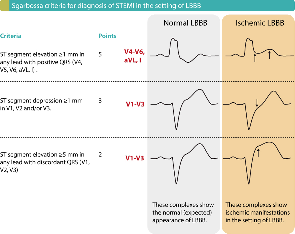
The ECG criteria for STEMI diagnosis was based on the European Society of Cardiology/ACCF/AHA/World Heart Federation Task Force for the Universal Definition of Myocardial Infarction as a new ST elevation at the J point in at least two contiguous leads of ≥2 mm (0.2 mV) in men or ≥ 1.5 mm (0.15 mV) in women in leads V2-V3 and/or of ≥1 mm.
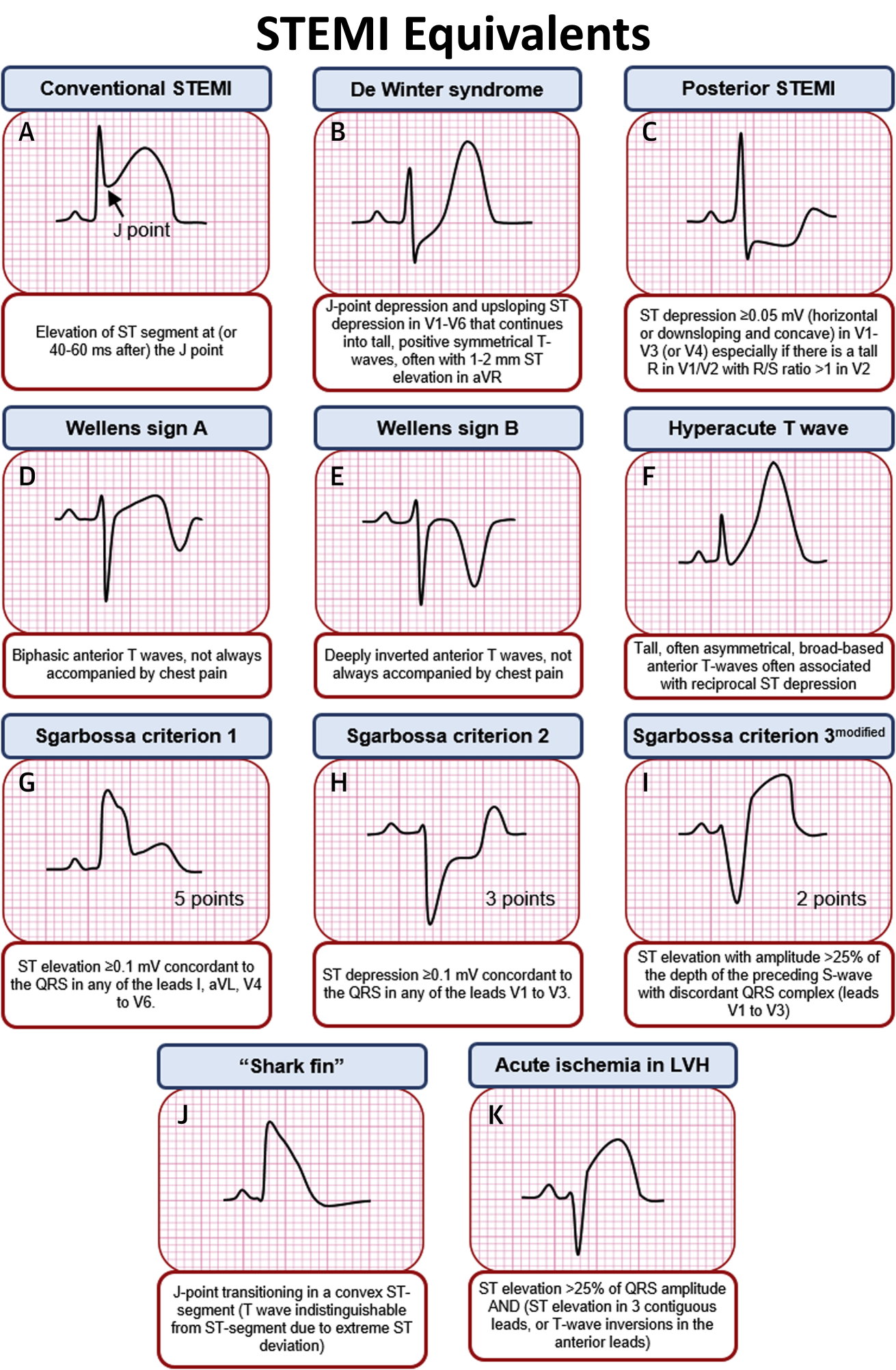
STEMI Equivalents on ECG • Conventional STEMI Elevation GrepMed
Infark miokard akut (IMA) masih merupakan salah satu penyebab kematian tertinggi di unit gawat darurat. Selama infark miokard akut yang disertai dengan ST elevasi (STEMI), elektrokardiogram (EKG) biasanya mengikuti perkembangan perubahan, dimulai dengan gelombang T hiperakut dan memuncak dengan elevasi segmen ST; gelombang Q patologis dapat muncul lebih awal dan/atau terlambat dalam prosesnya.
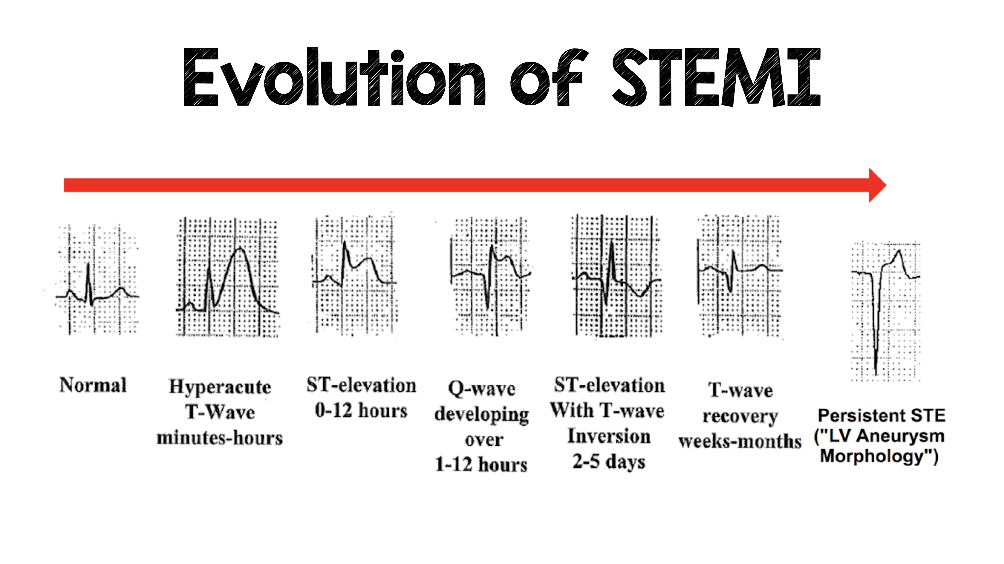
The Evolution of STEMI ECG Changes STEMI Evolution GrepMed
Consideration of typical EKG patterns in STEMI and STEMI mimickers. STEMI -EKG CRITERIA. •Diagnostic elevation (in absence of LVH and LBBB) defined as: - New ST elevation at J point in at least 2 contiguous leads -in leads V2-V3, men >2mm, women > 1.5mm -in other chest leads or limb leads, > 1mm. Alternative causes of ST-T changes.

Stemi Adalah Penyakit Homecare24
Kata Kunci: EKG, Evolusi, STEMI, TAVB ABSTRACT The electrocardiogram (ECG) modality is important for diagnosis and determining the course of ST-segment elevation myocardial infarction (STEMI). In STEMI there is an evolution or change in the ECG that indicates the course of the infarction. Without therapeutic intervention, the
STEMI (ST Elevation Myocardial Infarction) diagnosis, criteria, ECG & management ECG & ECHO
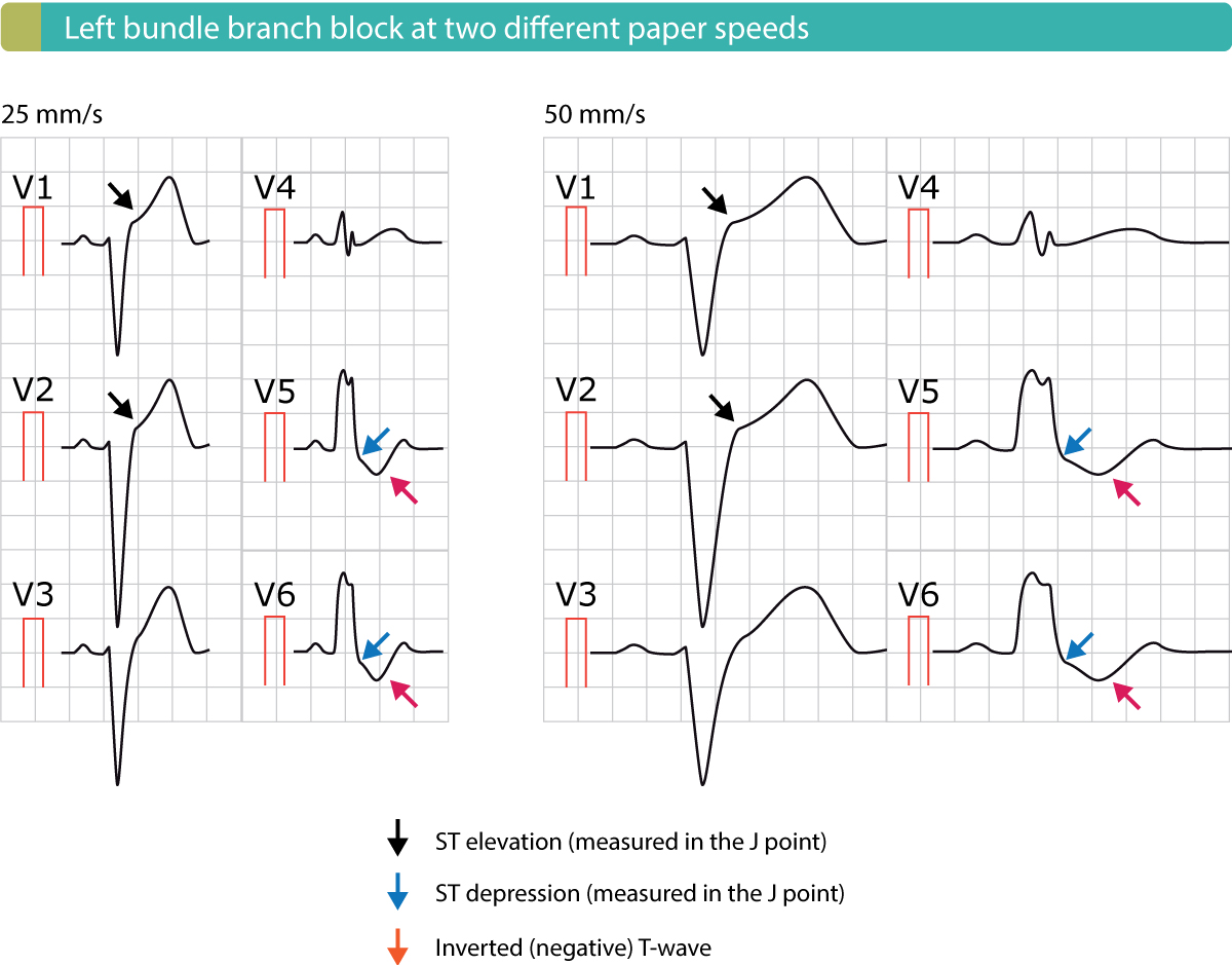
ECG (EKG) in acute STEMI (ST Elevation Myocardial Infarction) The ECG is the key to diagnosing STEMI. ECG criteria for STEMI are not used in the presence of left bundle branch block or left ventricular hypertrophy (LVH) because these conditions cause secondary ST-T changes which may mask or simulate ischemic ST-T changes. ST segment elevation is measured in the J-point and the elevation must.
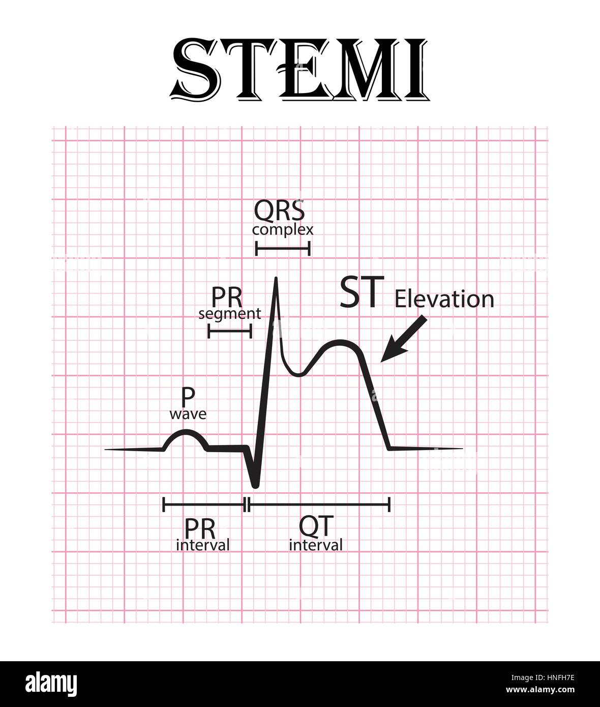
ECG of ST elevation myocardial infarction ( STEMI ) and detail of ECG ( P wave , PR segment , PR
Myocardial Ischaemia. Ed Burns and Mike Cadogan. Mar 16, 2022. Home ECG Library. This page covers the ECG signs of myocardial ischaemia seen with non-ST-elevation acute coronary syndromes (NSTEACS). ST-elevation and Q-wave myocardial infarction patterns are covered elsewhere: LMCA occlusion, Anterior STEMI, Lateral STEMI, Inferior STEMI, Right.
STEMI (ST Elevation Myocardial Infarction) diagnosis, criteria, ECG & management ECG learning
E-mail: [email protected]. ST-elevation myocardial infarction (STEMI) is a clinical syndrome defined by characteristic symptoms of myocardial ischemia in association with persistent electrocardiographic ST elevation (STE) and subsequent release of biomarkers of myocardial necrosis. 1 STE is the single best immediately available surrogate marker.

Mastering STEMI ECG
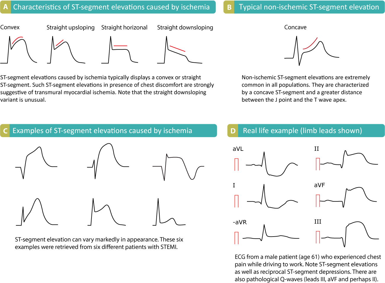
In STEMI/STE-ACS, on the other hand, reciprocal ST segment depressions are typical and there may be T-wave inversions in the same leads showing ST segment elevation. T-wave inversion may, however, occur in perimyocarditis, but only after normalization of the ST segment elevations (i.e these two ECG changes do not occur simultaneously).

Mastering STEMI ECG
Why is it called a STEMI? Myocardial infarction is the medical term for a heart attack. An infarction is a blockage of blood flow to the myocardium, the heart muscle. That blockage causes the heart muscle to die. A STEMI is a myocardial infarction that causes a distinct pattern on an electrocardiogram (abbreviated either as ECG or EKG).

STEMI and equivalent ECG Quiz
In patients with inferior STEMI, right-sided ECG leads should be obtained to screen for ST elevation suggestive of right ventricular (RV) infarction. (See Section 7.6.6 of the full-text guidelines and the ACC/AHA/ASE 2003 Guideline Update for the Clinical Application of Echocardiography.) (Level of Evidence: B)
Stadio intermedio posteriore STEMI (ECG) DocCheck
Specifically, In patients with STEMI, the presence and magnitude of microvascular obstruction was associated with the occurrence of mayor adverse cardiovascular events (adjusted HR 3.74 [95% CI 2.21-6.34]; P < 0.001). 47 Combining first pass myocardial perfusion and DE-MRI is also a unique means to visualize in one study session infarct size.

Evolution of Acute STEMI NCLEX Quiz
The evolution of ECG through these changes can occur rapidly after coronary artery occlusion. The emergency physician should be aware of the ECG findings that characterize the evolution of an STEMI with a sound understanding of the associated pathophysiology and clinical implication. This review discusses the changing ECG during an AMI.

Cómo reconocer un STEMI en un EKG de 12 derivaciones ECCtrainings
Continuous ECG-monitoring, including the need for monitoring staff, is potentially a significant contributor to costs of post-PCI care.. Older age, STEMI/NSTEMI, left main or multivessel disease, and presence of procedural complications are important risk factors for developing actionable arrhythmia alarms on cardiac telemetry. Presence of.
STEMI (ST Elevation Myocardial Infarction) diagnosis, criteria, ECG & management ECG & ECHO
A 12-lead ECG should be interpreted immediately (within 10 minutes) at first medical contact. ECG monitoring should start immediately and a defibrillator must be ready. Consider connecting leads V7, V8 and V9 in patients with high suspicion of posterior acute myocardial infarction (e.g due to reciprocal ST-segment depressions in V1, V2, V3).
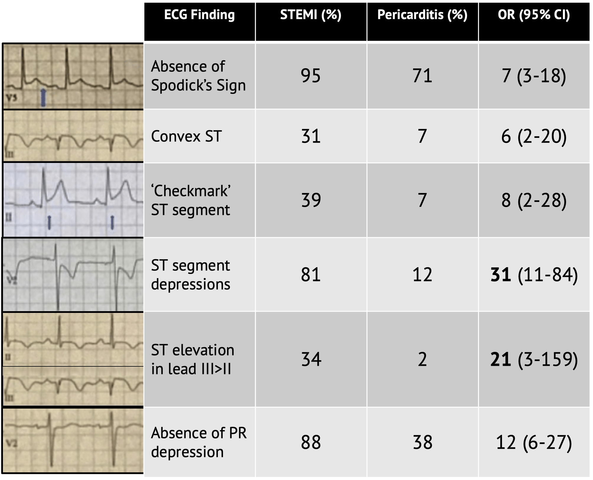
Differentiating STEMI from Pericarditis LaptrinhX / News
Anterior myocardial infarction carries the poorest prognosis of all infarct locations, due to the larger area of myocardium infarct size. A study comparing outcomes from anterior and inferior infarctions (STEMI + NSTEMI) found that compared with inferior MI, patients with anterior MI had higher incidences of: In-hospital mortality (11.9 vs 2.8%)
STEMI & NSTEMI A Nurse's Comprehensive Guide Health And Willness
Background: While ST-Elevation Myocardial Infarction (STEMI) door-to-balloon times are often below 90 min, symptom to door times remain long at 2.5-h, due at least in part to a delay in diagnosis. Objectives: To develop and validate a machine learning-guided algorithm which uses a single‑lead electrocardiogram (ECG) for STEMI detection to speed diagnosis.