Causes and Management of Wrist Joint Pain Complete Orthopedics
Penelitian ini bertujuan untuk mengetahui teknik pemeriksaan wrist joint proyeksi ulnar deviation pada kasus fraktur scaphoideus, dan untuk mengetahui perbedaan anatomi radiografi wrist joint proyeksi PA dengan ulnar deviation pada kasus fraktur scaphoideus.
Wrist Joint Anatomy Concise Medical Knowledge
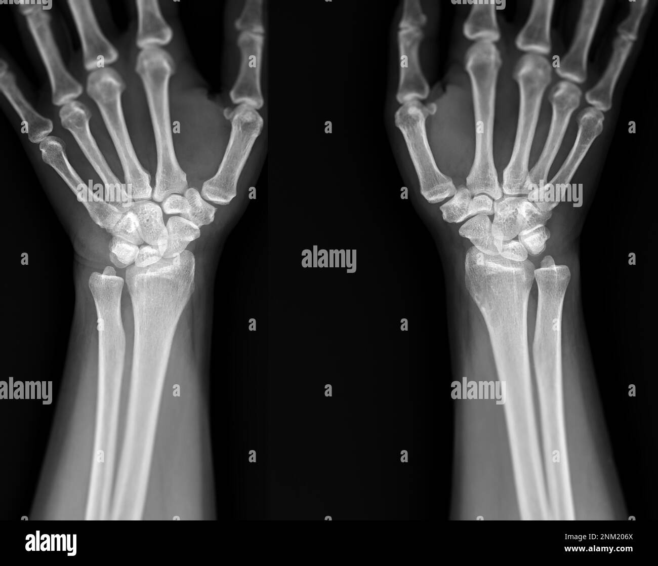
Menurut Merril's (2016), pada pemeriksaan wrist joint Anatomi radiografi wrist joint proyeksi PA ulnar deviation dengan proyeksi ulnar deviation yaitu CR 10-15 ° kearah chepalad tampak menunjukkan tampak tulang radius radiograf pada pertengahan film, distal dan ulna, carpal, dan tidak adanya rotasi pada pergelangan metacarpal proksimal.

Proyeksi Pemeriksaan Wrist Joint PDF
Teknik Pemeriksaan Radiografi Wrist Joint Dipos oleh Rini pada Mei 17, 2021 Proyeksi : PA Kaset : ukuran 18 x 24 cm kV : 60 ± 6 mAs : 4 FFD : 100 cm Posisi Pasien : Pasien duduk menyamping meja pemeriksaan,siku flexi 90°, posisi tangan dan lengan bawah berada di atas meja pemeriksaan
WRIST JOINT Samarpan Physiotherapy Clinic
Menurut jurnal dari Anil K. Bhat, dkk (2011) pemeriksaan Writ Jointcukup menggunakan proyeksi AP dan Lateral.Studi literatur ini bertujuan untuk mengetahui teknik pemeriksaan Wrist Joint dengan klinis dislokasi pada carpal dari beberapa jurnal literatur.
Wrist Joint Replacement (Wrist Arthroplasty) OrthoInfo AAOS
Abstract MRI is a medical imaging modality that works by utilizing hydrogen atoms in the body. Imaging that does not use ionizing radiation but uses an external magnetic field. MRI is able to.
Presentation1.pptx, ultrasound examination of the wrist joint.
Dari banyak indikasi pada wrist joint, beberapa indikasi yang dibahas pada penelitian ini adalah, fraktur, carpal tunnel syndrome dan pembedahan scapolunate. Penelitian ini bertujuan untuk mengetahui proyeksi yang tepat untuk mendiagnosa pada ke tiga indikasi tersebut.
Wrist Xray Interpretation OSCE Guide Geeky Medics
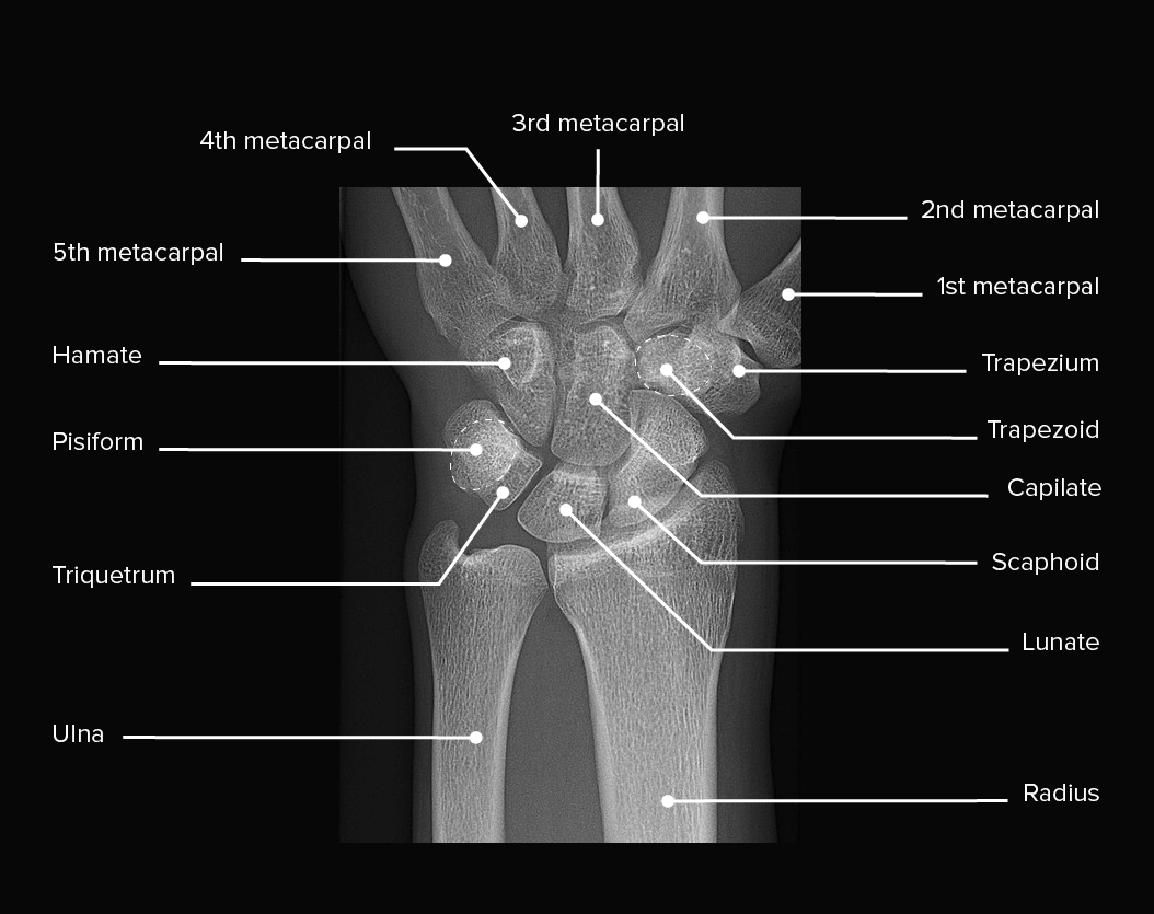
Untuk Proyeksi pemeriksaan Wrist Joint ada 8 yaitu : PA AP LATERAL BENDING ULNAR FLEXI BENDING RADIAL FLEXI PA OBLIQ AP OBLIQ KARPAL KANAL Tetapi untuk Proyeksi pemeriksaan di lapangan yaitu PA dan LATERAL untuk Proyeksi-proyeksi diatas bisa dilihat dari basic radiologi yang ada di buku. Proyeksi Pemeriksaan PA :
Wrist Joints Photograph by Asklepios Medical Atlas Pixels
TEKNIK RADIOGRAFI WRIST JOINT By yumanet Wednesday, April 18, 2018 Share Tweet Share Share Email. Radiografer. Persiapan Pasien Tidak memerlukan persiapan kusus, hanya melepas atau menyingkirkan benda yang dapat. Posisi Pasien : Duduk menyamping dari meja pemeriksaan . Posisi Objek : - lengan bawah menempel meja pemeriksaan

Wrist Joint AnatomyBones, Movements, Ligaments, Tendons Abduction, Flexion
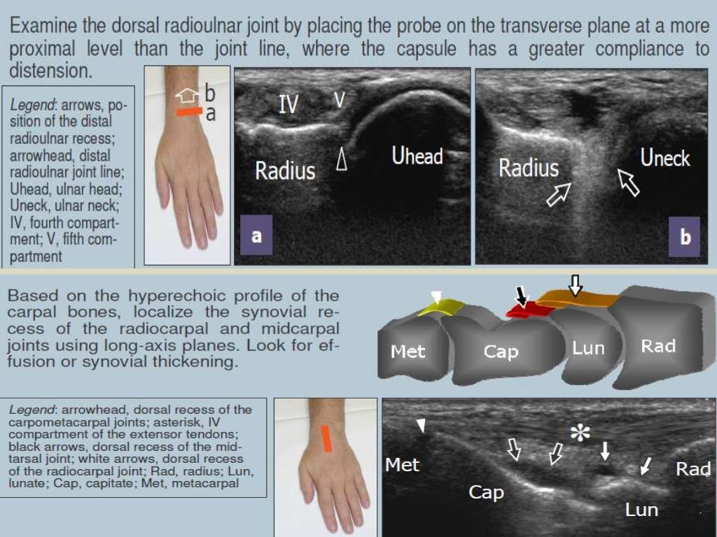
bringing the probe proximally, the distal radioulnar joint and midcarpal joints can be evaluated, in short and long axis, respectively. Ventral wrist. The hand is supinated for examination. evaluate the proximal carpal tunnel and distal carpal tunnel in short axis, with particular attention to the shape and echogenicity of the median nerve. The.

Wrist joint (Radiocarpal joint) Medically
And the reason for the wrist joint MRI examination in disruption of DRUJ case was added by the Fat Sat and STIR sequences, which were to suppress the fat around the wrist joint so the liquid odim or effusion fluid in the joint could be seen more clearly.. (2019). PROSEDUR PEMERIKSAAN MRI WRIST JOINT PADA KASUS DISRUPSI DISTAL RADIOULNAR.
Xray image of wrist joint front view of normal wrist joint Stock Photo Alamy
Conclusion : Procedure for MRI examination of wrist joints in disruption case of DRUJ using genu coil at Radiology Installation of Panti Rapih Hospital Yogyakarta.The reason for the wrist.
Wrist Joint Anatomy Concise Medical Knowledge
TEKNIK PEMERIKSAAN WRIST JOINT | PDF. Scribd adalah situs bacaan dan penerbitan sosial terbesar di dunia.
X Ray Wrist Joint Post Trauma Radiology Imaging
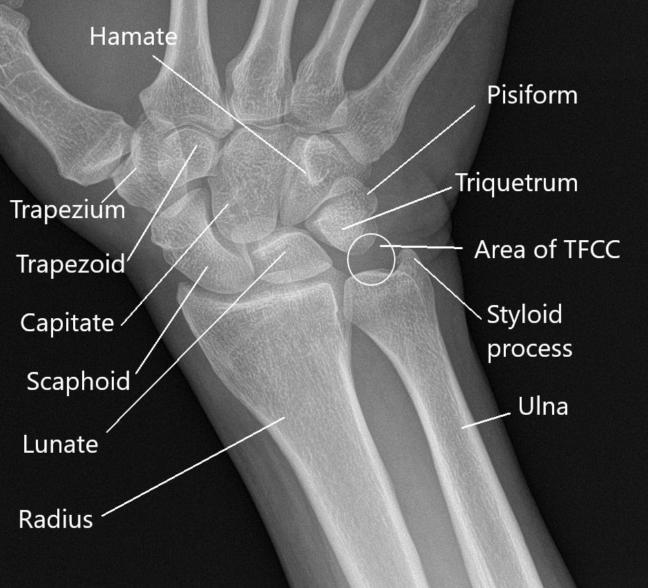
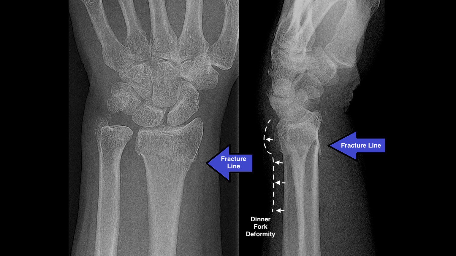
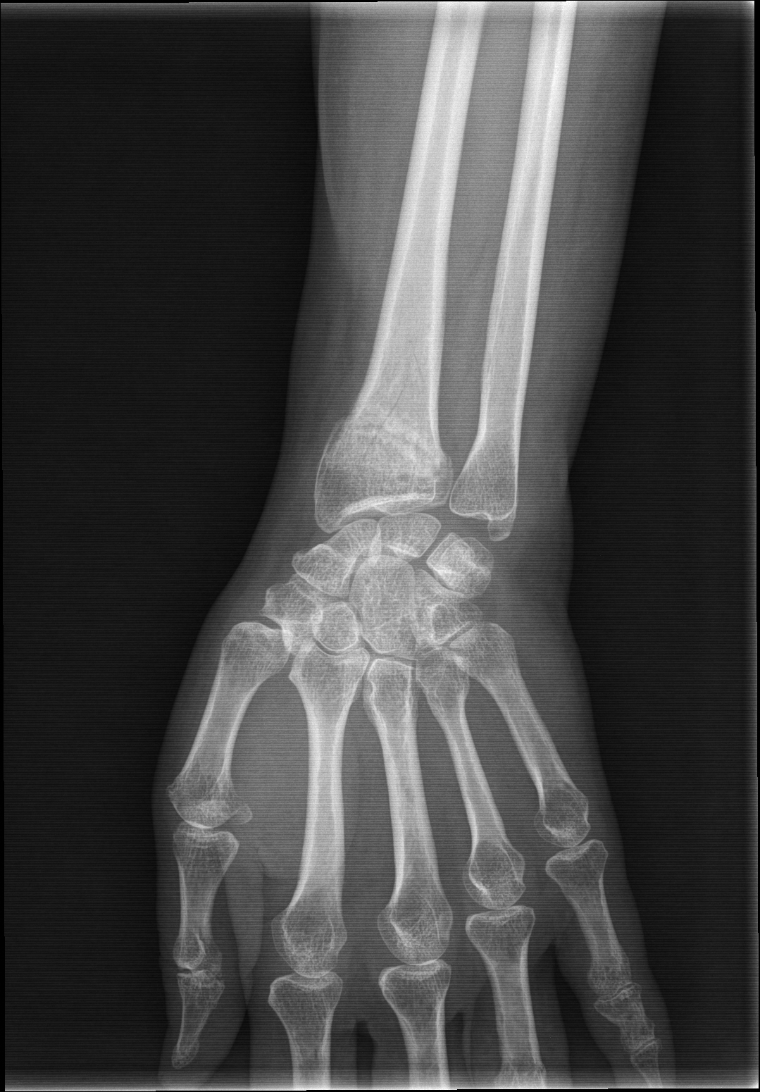
Abstract. Pemeriksaan radiografi wrist joint pada kasus fraktur scaphoid terdapat proyeksi khusus yaitu proyeksi PA ulnar deviation, pada posisi scaphoid yang superposisi dengan tulang trapezium, trapezoideum, dan lunatum akan mempengaruhi hasil radiograf, sehingga membutuhkan posisi dan arah sinar yang optimal dan dapat menampakkan tulang scaphoid.

Radiological imaging of the wrist joint Orthopaedics and Trauma
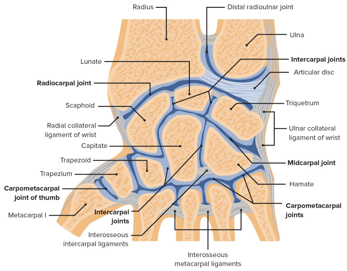
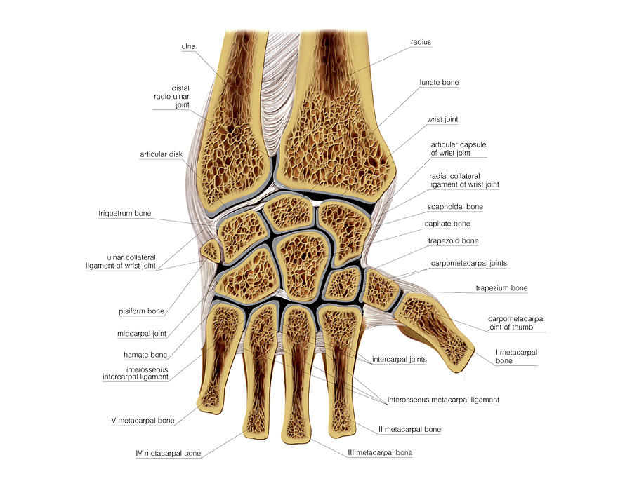
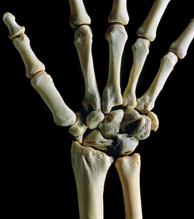
There are three joints in the wrist: Radiocarpal joint: This joint is where the radius, one of the forearm bones, joins with the first row of wrist bones (scaphoid, lunate, and triquetrum). Ulnocarpal joint: This joint is where the ulna, one of the forearm bones, joins with the lunate and triquetrum wrist bones.
Bones Of The Wrist Joint Photograph by James Stevenson/science Photo Library. Pixels
The CT hand and wrist protocol serves as an examination for the bony assessment of the wrist and is often performed as a non-contrast study and less often as a contrast-enhanced study. A CT wrist can be also conducted as a CT arthrogram for the evaluation of ligamentous injuries and the triangular fibrocartilage complex.. Note: This article aims to frame a general concept of a CT protocol for.
Wrist Joint Movements by Maurizio De Angelis/science Photo Library
High field imaging. MR imaging of the wrist and elbow is now commonly performed at intermediate field strengths of 1.5T or higher. Imaging at 3.0T has become increasingly common for clinical evaluation, while even higher field systems (7.0T) are being evaluated in the research realm 9.While initially used for neurological imaging, numerous studies have confirmed the benefits and abilities of.