
BiRADS for Mammography and Ultrasound 2013 Mammography, Mammogram, Ultrasound
USG mammae memiliki biaya yang bervariasi, dimulai dari Rp. 250.000 hingga lebih dari Rp. 750.000 tergantung dari rumah sakit. Apa Itu USG Mammae? Prosedur pencitraan dengan menggunakan teknologi gelombang suara berfrekuensi tinggi untuk memproduksi gambar payudara bagian dalam dengan menggunakan alat sensor ( probe ) dan ditampilkan melalui.

USG Mammae dan Penjelasannya Bunda Denpasar
Epidemiology. Breast cancer is the most common nonskin malignancy in women. In the affluent populations of North America, Europe, and Australia, 6% of women develop invasive breast cancer before age 75, compared to a 2% risk in developing regions of Africa and Asia 8.The difference has been attributed to risks associated with a Westernized lifestyle, including high-calorie diet rich in fat and.
What Does Breast Cancer Look Like On An Ultrasound
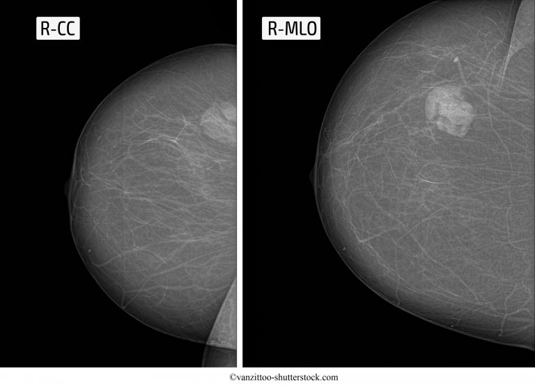
Breast ultrasound is an important modality in breast imaging. It is the usual initial breast imaging modality in those under 30 years of age in many countries ref. In assessing for malignancy, it is important to remember that one must use the most suspicious feature of three modalities (pathology, ultrasound and mammography) to guide management.

Proses USG Payudara Biaya usg payudara USG Mammae with doppler Deteksi tumor kanker
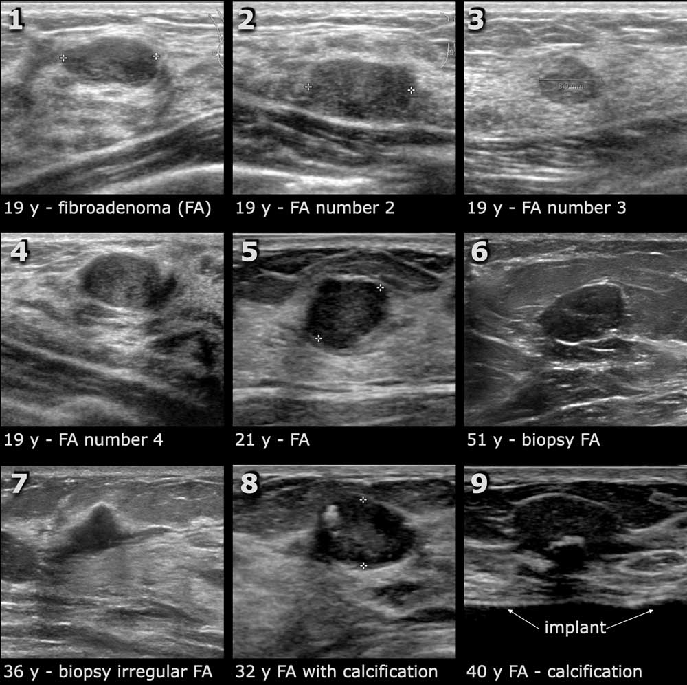
FIBROADENOMA is the most common breast tumor in adolescent girls and women younger than 25 years. Although the peak incidence is between the second and third decades of life, it is not uncommon in postmenopausal women, with an increased incidence after hormone replacement therapy. 1 Overall, it occurs in approximately 10% of women and accounts for about 50% of breast biopsies performed. 2

Cek Kondisi Payudara dengan USG Mammae di RSIA Nuraida Bogor
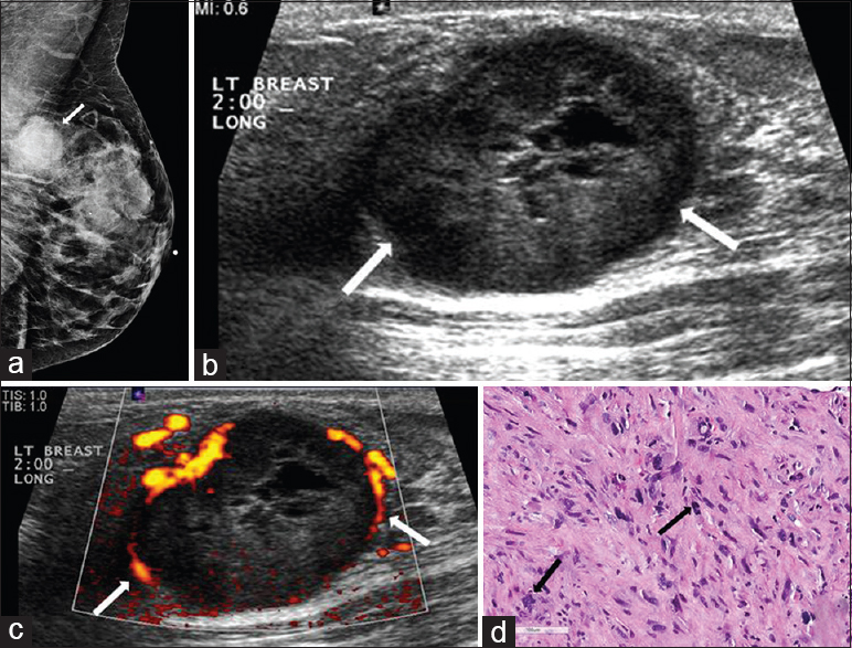
Phyllodes tumors are very similar to intracanalicular fibroadenomas, and histological underestimation is possible when a limited amount of sampling material is available (e.g. cytological sampling but also core biopsy). In these cases, diagnosis is assumed when the nodules are larger than 3 cm in diameter or fast-growing (benign lesions have an.
Package USG Mammae Mandaya Hospital Group
It is a common 'normal' finding, that is seen in 55% of men at autopsy. The peak incidence is 60 - 69 years. It is significant if it is new or symptomatic. In elderly males gynecomastia makes up 65% of all breast lesions. 25% is carcinoma and 10% are other lesions. Mammogran and rotated ultrasound image.

Phyllodes Tumors of the Breast Ultrasonographic Findings and Diagnostic Performance of
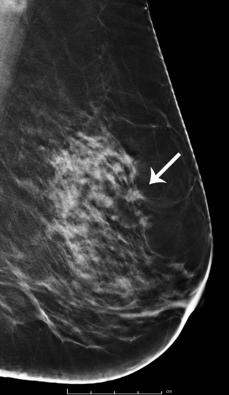
Breast cancer is the most common malignant tumor in women. A woman's risk of getting breast cancer increases with age. Most women diagnosed with breast cancer are over the age of 50, but younger women can also get breast cancer. The first noticeable clinical symptoms are: lump or area of thickened breast tissue; retraction of the nipple

Mammogram radio imaging for breast cancer diagnosis ODC
Paket USG Mammae Cegah Kanker Payudara adalah deteksi dini kanker payudara yang paling efektif & cepat untuk mengetahui kanker/tumor payudara. Skip to content. Mandaya Hospital Group. A Hospital Like No Other. Mandaya Royal Puri +62-21-5092-8888 +62-811-1900 2000 . Mandaya Karawang
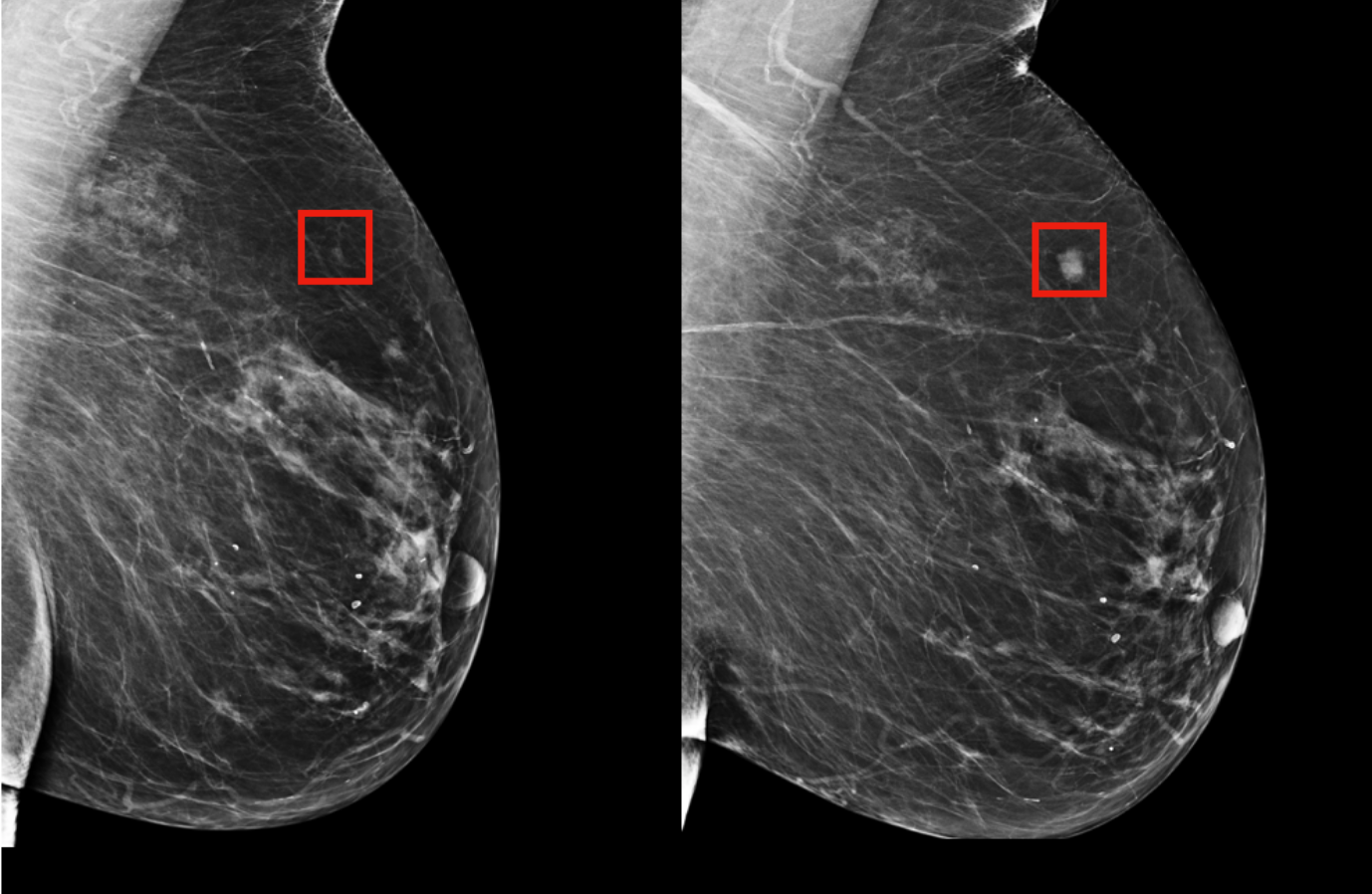
Breast Cancer Screening with 3D Mammography or Tomosynthesis Radiology & Imaging, MA, CT
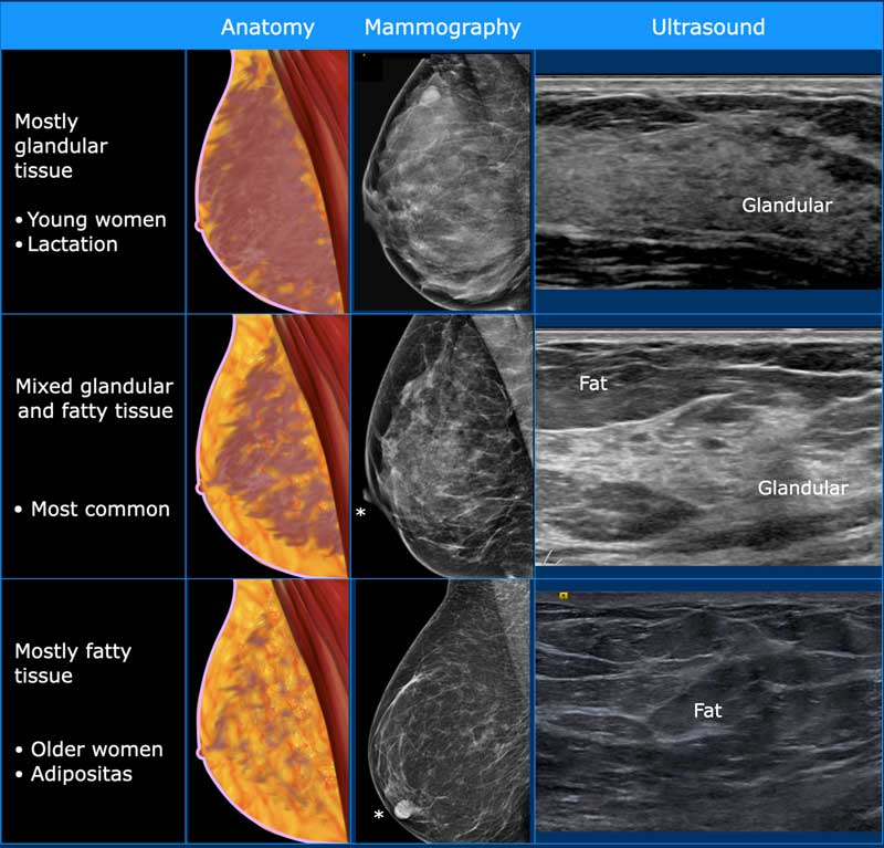
USG Mammae sering digunakan sebagai pelengkap atau pemeriksaan tambahan selain mamografi, yang merupakan pemeriksaan pencitraan standar untuk deteksi dini kanker payudara. Pemeriksaan ini dapat memberikan informasi tambahan atau membantu mengklarifikasi temuan yang ditemukan melalui mamografi atau pemeriksaan fisik.

Dikira Hamil di Luar Nikah Usai Unggah Hasil USG, Kesha Ratuliu Angkat Bicara Itu Adalah Tumor
Breast ultrasound has developed into a practical solution for the evaluation of breast disease. Although mammography remains the gold standard for breast cancer screening, it presents certain imaging limitations with dense breast parenchyma. Due to this, ultrasound and magnetic resonance imaging (MRI) have been expanding their role as part of supplementary breast screening procedures.[1]
Rare Malignant Tumors of the Breast Journal of Clinical Imaging Science
Persiapan sebelum USG mammae. Sebenarnya tidak ada persiapan khusus sebelum melakukan USG payudara. Namun, sebaiknya Anda memerhatikan hal di bawah ini untuk memudahkan selama pemeriksaan dan mendapat hasil yang optimal. Jangan mengoleskan losion, krim, bubuk, atau produk skin care maupun riasan apapun ke area kulit payudara.
Fibroadenoma da mama, sintomas, tratamento e prognóstico
Ultrasound. On ultrasound, mucinous carcinomas often display mixed echogenicity with mixed solid and cystic components. Posterior acoustic enhancement is common. At times the lesion can be isoechoic to breast tissue which can make diagnosis difficult 3. Distal enhancement and microlobulated margins are also commonly found in mucinous carcinomas.
Using AI to predict breast cancer and personalize care MIT News Massachusetts Institute of
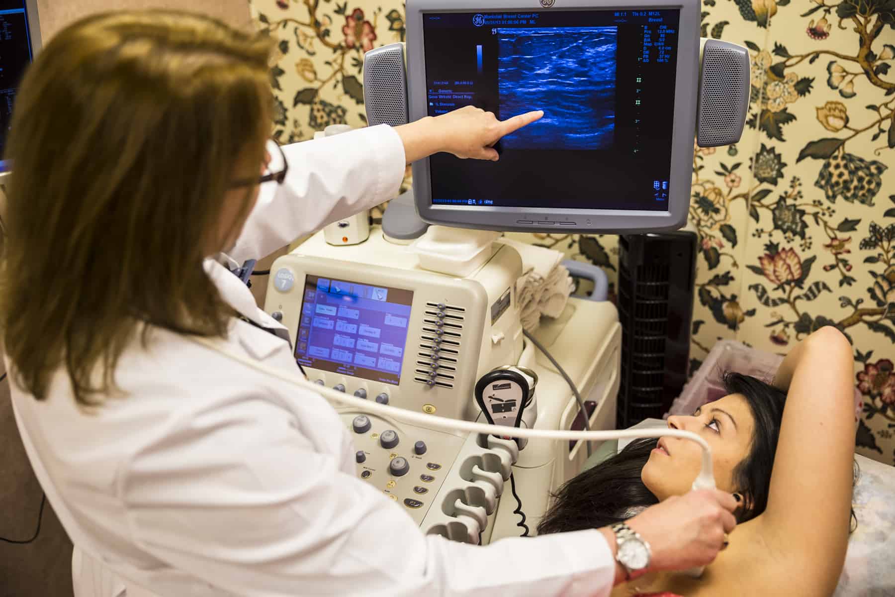
USG Mammae. A Mammae USG or breast ultrasound is one type of ultrasound that examines the condition of the breast and detects disorders and various forms of abnormalities in the breast, such as cysts and tumors. Mammary ultrasound works using high-frequency sound waves or ultrasound. The USG's wave will appear from the scanner machine that.
Ultrazvuk prsníka potrebné funkcie, postupy a príprava
Phyllodes tumor of the breast is a rare, yet clinically significant, fibroepithelial neoplasm accounting for 1% of all breast neoplasms [].Women classically present with a rapidly growing palpable abnormality that triggers a diagnostic imaging workup [].Phyllodes tumors are biphasic, composed of both epithelial and stromal components [], and have a characteristic leaflike architecture with.

Imaging features of breast cancer with marked hemosiderin deposition A case report European
Ultrasound can also be used to help guide a biopsy needle into an area of the breast so that cells can be taken out and tested for cancer. This can also be done in swollen lymph nodes under the arm. Ultrasound is widely available and is fairly easy to have done, and it does not expose a person to radiation. It also tends to cost less than other.
What Does Breast Cancer Look Like On An Ultrasound
Epidemiology. Phyllodes tumors account for less than 0.3-1% of all breast neoplasms 13. It is predominantly a tumor of adult women, with very few examples reported in adolescents. The occurrence is most common between the ages of 40 and 60, before menopause (peak incidence ~45 years). This is about 15 years older than the typical age of.
